The University of British Columbia
UBC - A Place of Mind
The University of British Columbia
Faculty of MedicineProgram in Cryo-EM Guided Drug Design
  • Home
  • Research
  • Publications
  • News
  • Team
  • Contact
  • Imaging Proteins Using Electrons

    Imaging with electrons allows researchers to discern atomic details of large, complex molecules like proteins. During the imaging process, the interaction of electrons with the molecules being imaged inflicts damage that can alter the structure. Cryogenic temperatures (cryo-EM) can reduce the extent of this damage, but as we showed in a recent study, some parts […]

  • Visualizing Proteins with Cryo-EM

    This image displays a number of the critical elements for determining the structure of a protein by cryo-electron microscopy. First, a solution containing many identical copies of a protein are spread on a grid (upper left), and quickly frozen to liquid nitrogen temperatures. These are imaged with an electron beam (teal), producing black-and-white images showing […]

  • HIV in the Brain

    The human immunodeficiency virus (HIV) primarily attacks helper T cells, rapidly-dividing cells that are critical components of the immune system. The immune system keeps the virus in check by eliminating infected T cells. However, the virus can also occasionally invade long-lived cells like macrophages in the blood, or astrocytes in the brain, hiding away and […]

  • T cells Transfer HIV

    This post is dedicated to the memory of our colleague Gavin Murphy, the primary author of this study, who passed away July 4, 2017. The human immunodeficiency virus (HIV) is the causative agent for AIDS (or Acquired Immune Deficiency Syndrome). The virus primarily infects CD4+ T cells, a class of immune cells critical for defense […]

  • Stepping into a Melanoma Cell

    The image above shows the internal organization of a melanoma cell, a cancer cell descended from pigment-containing skin cells. This image was generated from data acquired with focused ion beam scanning electron microscopy (FIB-SEM), a method for 3D imaging of cells that we have been developing in our laboratory for several years. In a study […]

  • Formation of the HIV Core

    Before reaching their mature, fully infectious form, HIV virus particles transition through an immature, non-infectious state. In the non-infectious form, the inside of the virus particle is lined with a lattice of Gag proteins. During the virus’ transition from the immature state to the fully infectious state, a piece of the Gag protein (the capsid) […]

  • A 3D View of SIV

    The simian immunodeficiency virus (SIV), like its close relative human immunodeficiency virus (HIV), infects immune cells (such as T cells) using its envelope glycoprotein spike. Many copies of this spike dot the membranous outer surface of the virus, while the virus’ genetic material is packed within, inside a conical core. Individual copies of the virus, […]

  • Dendritic Cell with HIV

    Classic images of dendritic cells show long finger-like appendages flowing around and circling the cell body; these appendages, called dendrites, are the feature that gave rise to the cell type’s name. But in studies published by our lab in 2010-2011, new high-resolution 3D images revealed that the appendages are more like flaps than fingers, and […]

  • Visualizing Ebola Virus

    As with most viruses, the Ebola virus itself is very small – too small to be seen even in a high-powered light microscope. But when visualized in an electron microscope, the surface of the filament-shaped virus particle (also known as a virion) can be seen, studded with small proteins on its surface (upper right of […]

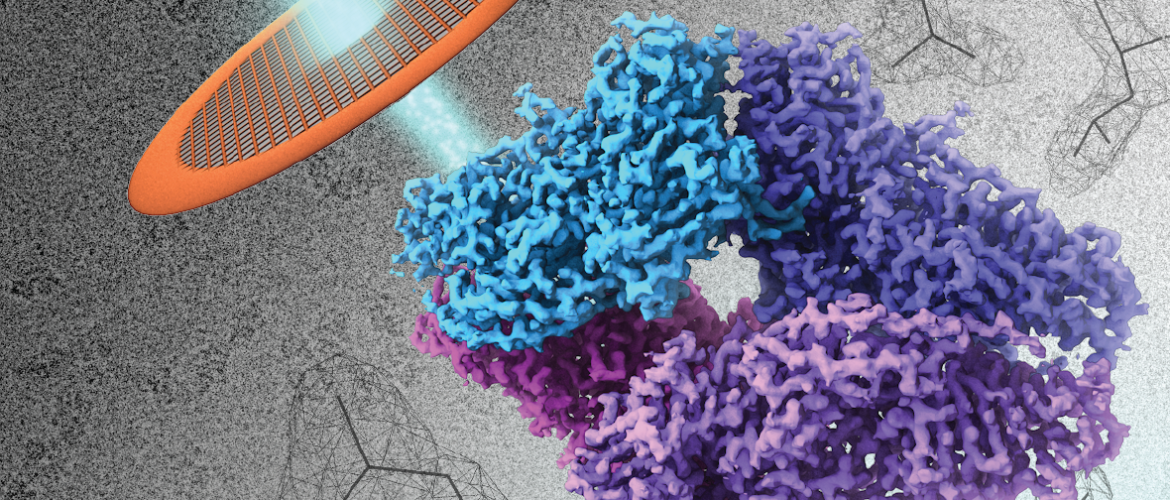
Cryo-electron microscopy (Cryo-EM) and drug design

Our research program is focused on exploring frontiers in structural biology and drug design using cryo electron microscopy (cryo-EM), with the central goal of accelerating the development of effective therapeutic agents. The program is guided by two overarching themes:
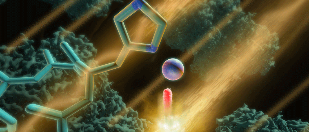
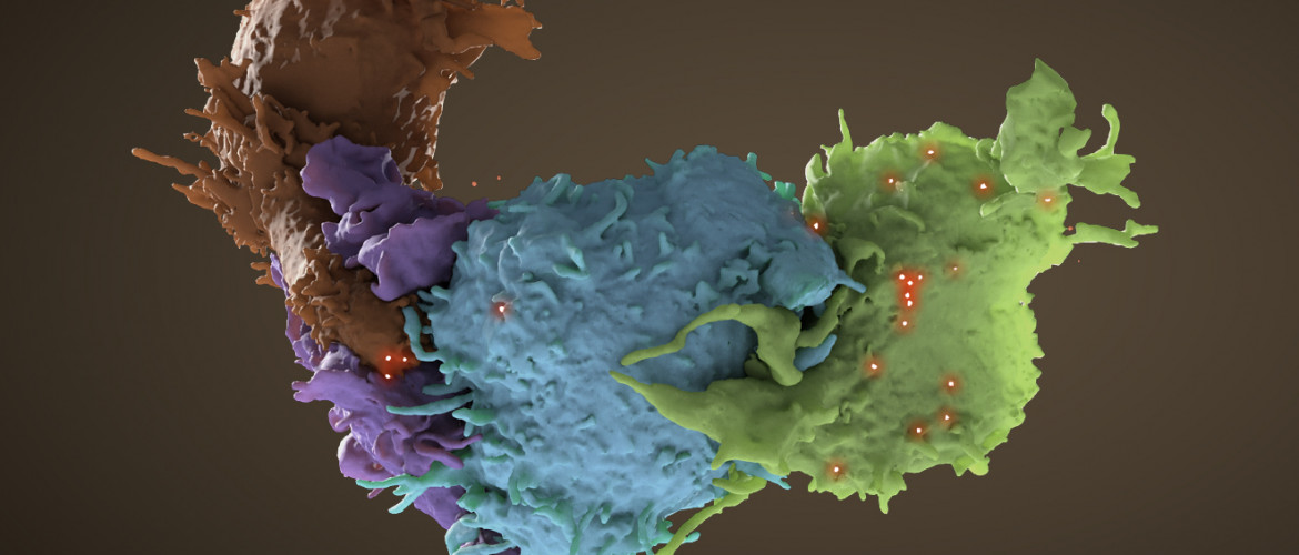
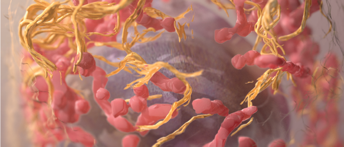
Drug Design

A highly focused team effort to use high-resolution cryo-EM imaging of native protein complexes to design effective therapeutic agents targeting key drivers of diseases such as cancer, infectious diseases and brain disorders.

Technology Development

An interdisciplinary approach towards developing next generation technologies in cryo-EM and related methods using automated workflows coupled with advanced image processing and machine learning.

  • News
UBC Medicine on Twitter

Twitter | February 22, 2023

We were delighted to welcome Minister Selina Robinson for a tour of UBC’s Cryo-EM Guided Drug Design lab, where UBC researchers and students are accelerating the process of drug discovery.

Selina Robinson on Twitter

Twitter | February 21, 2023

Thank you to @UBC & @cryoem_UBC for showing me the great work of students and faculty with the cryo electron microscope. So fascinating!

More news
Program in Cryo-EM Guided Drug Design
Faculty of Medicine
Djavad Mowafaghian Centre for Brain Health
Room 3404 – 2215 Wesbrook Mall
Vancouver, BC Canada V6T 1Z3
Tel 604 822 8073
Website electron.med.ubc.ca
Email cryoem.lab@ubc.ca
Find us on
  
Back to top
The University of British Columbia
  • Emergency Procedures |
  • Terms of Use |
  • Copyright |
  • Accessibility