Image credit: Veronica Falconieri, National Cancer Institute
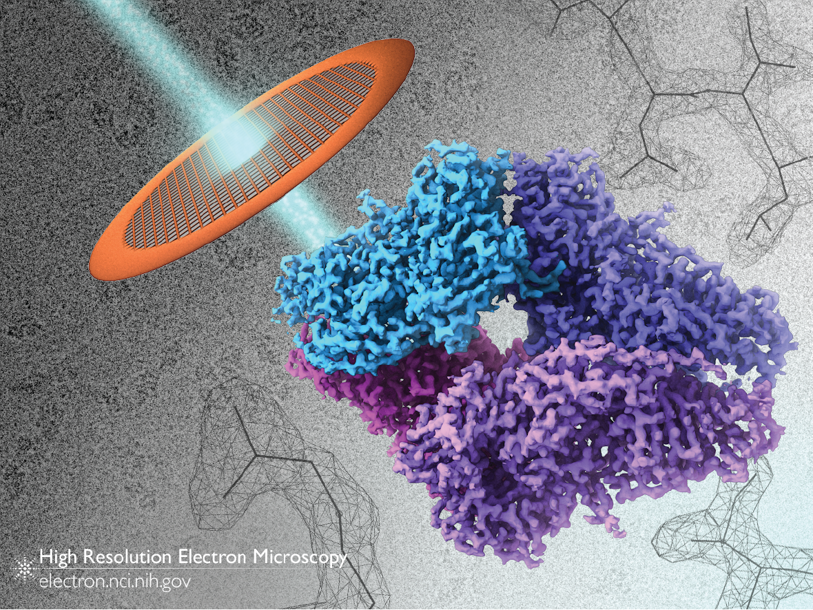
This image displays a number of the critical elements for determining the structure of a protein by cryo-electron microscopy. First, a solution containing many identical copies of a protein are spread on a grid (upper left), and quickly frozen to liquid nitrogen temperatures. These are imaged with an electron beam (teal), producing black-and-white images showing “silhouettes” of proteins (background, upper left). Thousands of these images are computationally combined to produce a 3D “map”, a graphical representation of the protein’s shape. A high resolution map of β-galactosidase is shown in the lower right. Given sufficient resolution, a chain of amino acids can be threaded through the map, creating an atomic model of the protein’s structure (background, upper right).
This process is shown in a video that accompanied our 2015 publication of a 2.2 Å resolution map of β-galactosidase.
Full-size image: Download (2.5MB)
Related Reference: Bartesaghi A, Merk A, et al. 2.2 Å resolution cryo-EM structure of β-galactosidase in complex with a cell-permeant inhibitor. Science. 2015 Jun 5;348(6239):1147-51. doi: 10.1126/science.aab1576.