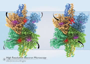
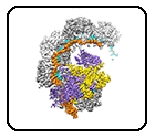
Two States of Glutamate Dehydrogenase
The enzyme glutamate dehydrogenase (GDH) functions as a critical part of the metabolic machinery of most cells. This enzyme converts the protein building-block glutamate to α-ketoglutarate, with the help of the small molecule “co-enzyme” NADH. During its catalytic cycle, GDH shifts from an “open” conformation (the catalytic pocket, with binding sites for glutamate and NADH, […]
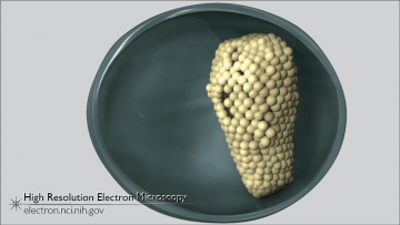
Formation of the HIV Core
Before reaching their mature, fully infectious form, HIV virus particles transition through an immature, non-infectious state. In the non-infectious form, the inside of the virus particle is lined with a lattice of Gag proteins. During the virus’ transition from the immature state to the fully infectious state, a piece of the Gag protein (the capsid) […]
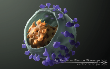
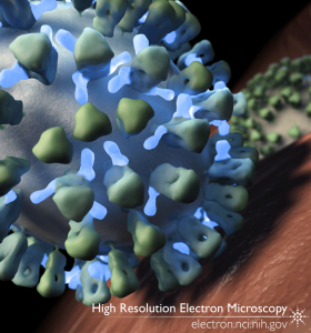
A 3D View of SIV
The simian immunodeficiency virus (SIV), like its close relative human immunodeficiency virus (HIV), infects immune cells (such as T cells) using its envelope glycoprotein spike. Many copies of this spike dot the membranous outer surface of the virus, while the virus’ genetic material is packed within, inside a conical core. Individual copies of the virus, […]
A Wrench in the Works
This post is dedicated to the memory of our colleague Soojay Banerjee, the primary author of this study, who passed away October 24, 2017. While creating new proteins is critical for a cell, getting rid of excess or unneeded proteins is equally critical. This is particularly true for cancer cells, which tend to have higher […]
Microbiology catches the cryo-EM bug
Earl L, Falconieri V, Subramaniam S. Current Opinion in Microbiology 43: 199-207 (2018)
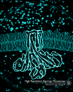
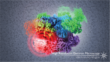
CorA Magnesium Channel
Charged ions – like magnesium, chloride, sodium, potassium, and others – are used by cells to make proteins and other building blocks, to pass signals within the cell and to other cells, and for a variety of other purposes. Maintaining just the right amount of these ions inside the cell requires the action of highly […]
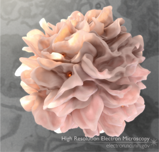
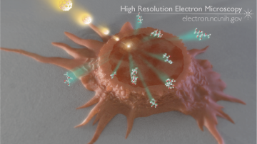
Dendritic Cell with HIV
Classic images of dendritic cells show long finger-like appendages flowing around and circling the cell body; these appendages, called dendrites, are the feature that gave rise to the cell type’s name. But in studies published by our lab in 2010-2011, new high-resolution 3D images revealed that the appendages are more like flaps than fingers, and […]
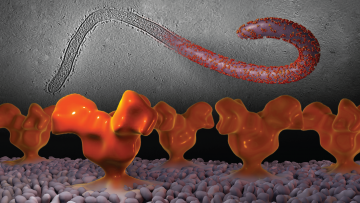
Visualizing Ebola Virus
As with most viruses, the Ebola virus itself is very small – too small to be seen even in a high-powered light microscope. But when visualized in an electron microscope, the surface of the filament-shaped virus particle (also known as a virion) can be seen, studded with small proteins on its surface (upper right of […]
Inhibiting Influenza
The surface of the influenza (flu) virus is covered with two proteins: hemagglutinin (HA), and neuraminidase (NA). While the immune system can recognize both surface proteins, because HA is the protein that enables viral entry into cells, it is a high priority target for researchers trying to create a long-lasting, universal flu vaccine. The HA […]
Chemical Imaging
What if you could point at a specific spot in a cell or tissue and know precisely what molecules were there, irrespective of labeling, fluorescence or chemistry? In an article published in the January 26, 2011, edition of Analytical Chemistry, Szakal and colleagues demonstrated that mass spectrometry, paired with a focused beam of gallium ions, […]