Image credit: Veronica Falconieri, National Cancer Institute
Charged ions – like magnesium, chloride, sodium, potassium, and others – are used by cells to make proteins and other building blocks, to pass signals within the cell and to other cells, and for a variety of other purposes. Maintaining just the right amount of these ions inside the cell requires the action of highly specific channels – small pores that open and close according to specific signals, and which allow just one type, or select types, of these ions to pass through.
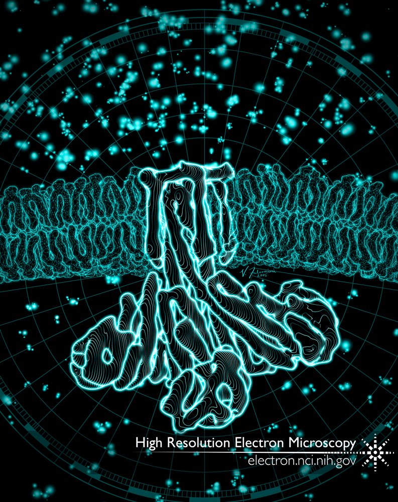
CorA is an ion channel found in prokaryotic cells. This channel senses the concentration of magnesium inside the cell, and when it gets too low, opens to allow more magnesium in from the outside. When the channel is closed, the part of the channel facing into the cell looks like a symmetric, 5-pointed star. This star-like arrangement is stabilized by magnesium ions, which bind and hold together each lobe (or “point”) of the star with the one next to it.
In a study published in early 2016, we used cryo-EM to study the structure of CorA when the magnesium concentration is low. Our structures showed that the five lobes are no longer arranged symmetrically. One slips outwards away from the center of the star, and then two on the opposite side fall in; this movement opens the pore at the membrane, wide enough that magnesium ions can flow in.
This journal cover concept art shows a side-view of the structure of CorA in the open state, with the pore of the channel buried in a lipid bilayer. Greater concentrations of magnesium (the blue dots) are found outside the cell (top) than inside (bottom).
Full-size image: Download (0.4MB)
Related Reference: Matthies D, Dalmas O, et al. Cryo-EM structures of the magnesium channel CorA reveal symmetry break upon gating. Cell. 2016 Feb 11;164(4):747-56. doi: 10.1016/j.cell.2015.12.055.