Image credit: Thao Do & Subramaniam Lab, National Cancer Institute
The human immunodeficiency virus (HIV) primarily attacks helper T cells, rapidly-dividing cells that are critical components of the immune system. The immune system keeps the virus in check by eliminating infected T cells. However, the virus can also occasionally invade long-lived cells like macrophages in the blood, or astrocytes in the brain, hiding away and re-emerging at later times to infect new helper T cells. The long-lived cells can form a “reservoir” that allows the virus to evade anti-viral treatment.
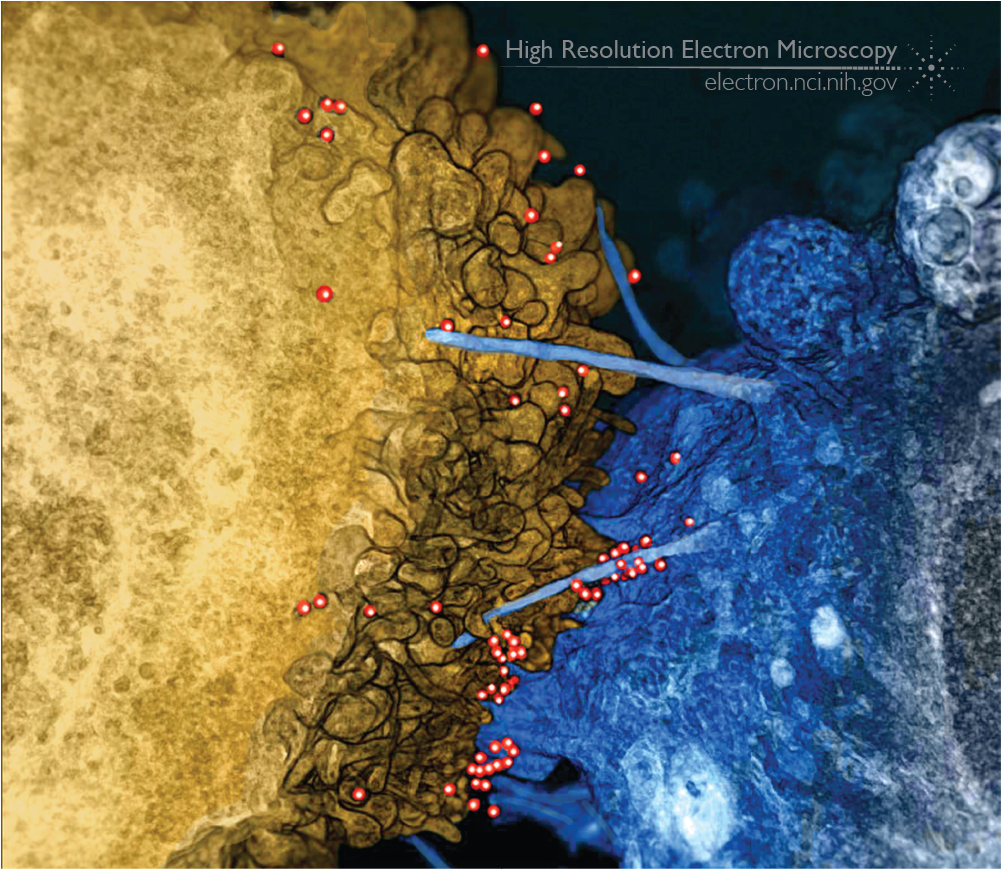
In a collaboration with the laboratories of Avi Nath (NINDS) and Jeff Lifson (NCI/Leidos), published in 2014, we used focused ion beam scanning electron microscopy (FIB-SEM) to visualize contacts between HIV-infected T cells and uninfected astrocytes at high resolution in 3D. Astrocytes, a type of support cell in the brain, send out long membrane extensions that generally interact with other cells. We found that infected T cells attached to astrocytes, making long-lived connections. At high resolution, the astrocytes formed thin, membranous bridges across to the T cell, with HIV particles clustered on these bridges and crossing over from the T cell to the astrocyte.
The image above shows a colorized version of the 3D reconstruction of our FIB-SEM data. On the right, the astrocyte (in blue) has long filopodial extensions crossing towards the T cell on the left (in gold). HIV particles (red) are present on the surface of the T cell, on the filopodial bridges, and on the surface of the astrocyte.
Full-size image: Download (1.4MB)
Related Reference: Do T, Murphy G, Earl LA, Del Prete GQ, Grandinetti G, Li G, Estes J, Rao P, Trubey C, Thomas J, Spector J, Bliss D, Nath A, Lifson JD, and Subramaniam S. 3D imaging of HIV-1 virological synapses reveals membrane architectures involved in virus transmission. J Virol. 2014 Sep;88(18):10327-39. doi: 10.1128/JVI00788-14. Epub 2014 Jun 25.