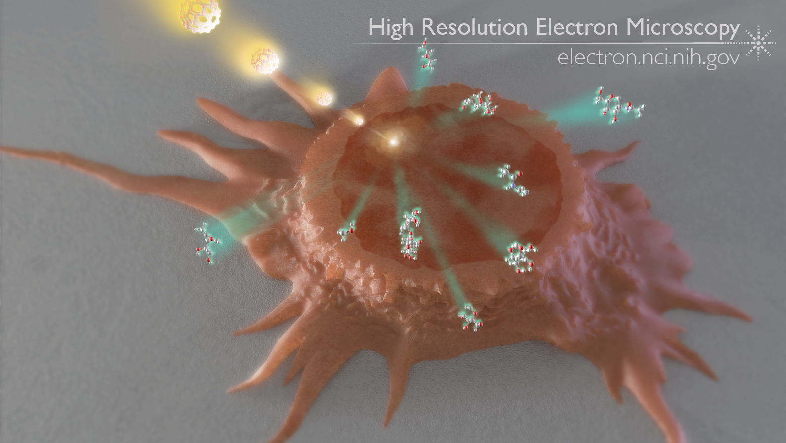
Image credit: Donald Bliss, National Library of Medicine
What if you could point at a specific spot in a cell or tissue and know precisely what molecules were there, irrespective of labeling, fluorescence or chemistry? In an article published in the January 26, 2011, edition of Analytical Chemistry, Szakal and colleagues demonstrated that mass spectrometry, paired with a focused beam of gallium ions, can do exactly that.
The technique is a special version of secondary ion mass spectrometry (SIMS). In this technique, a freeze-dried sample of cells or tissue is abraded with a beam of ions. Ordinary SIMS uses a laser beam; in the technique pioneered in this study, a gallium ion beam is used instead. The gallium ions, focused on a specific “pixel”, or surface area, scatter molecules off the surface of the tissue. These molecules then fly through a vacuum towards a detector, which records the mass of each molecule. From the mass, one can then determine what types of molecules were present in that “pixel” of the tissue or cell.
Although more work needs to be done to refine the technique, SIMS paired with a gallium ion beam has the potential to create a 3D representation of the chemical composition of a sample of cells or tissue, something that neither imaging nor standard SIMS can accomplish.
Full-size image: Download (1.6MB)
Related Reference: Szakal C, Narayan K, et al. Compositional Mapping of the surface and interior of mammalian cells at submicrometer resolution. Anal Chem. 2011 Feb 15;83(4):1207-13.