Gram-negative bacteria, like Escherichia coli and Salmonella species, can wreak havoc on their human hosts when they develop antibiotic resistance. But even bacteria that can chuck out antibiotics can’t defend against this tiny hunter, the small prokaryote Bdellovibrio bacteriovorus.
Like its prey, Bdellovibrio is also a gram-negative bacterium. It can replicate either on its own or inside another bacterium, harvesting the DNA, proteins, and lipids of its prey. But much about Bdellovibrio remains a mystery: despite a complete genome sequence of this species, we still don’t understand how Bdellovibrio divides, nor do we understand the packing and regulation of Bdellovibrio’s genetic material.
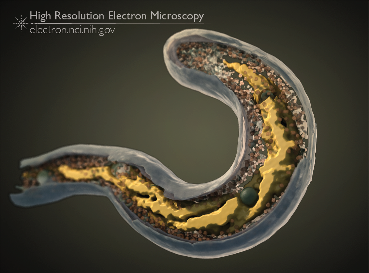
This image, from an article published in 2011, shows the 3D structure of the Bdellovibrio nucleoid, a structure made up of the bacterium’s genome. We used cryo-electron tomography, a technique that can visualize whole small bacteria like Bdellovibrio in great detail in 3D. For wild-type, normal Bdellovibrio bacteria, we saw either a tightly packed, amorphous nucleoid, or a nucleoid organized into two twisted strands.
In other gram-negative species, the organization of the nucleoid is managed by the actin-like molecule MreB. When we imaged Bdellovibrio bacteria carrying MreB tagged with a fluorescent molecule, we saw that this altered MreB disrupted cellular division, creating cells that were longer, and displayed more obviously helical nucleoids (shown in the image above).
Full-size image: Download (1.2MB)
Related Reference: Butan C, Hartnell LM, et al. Spiral architecture of the nucleoid in Bdellovibrio bacteriovorus. J Bacteriol. 2011 Mar;193(6):1341-50.