Image credit: Donald Bliss, National Library of Medicine
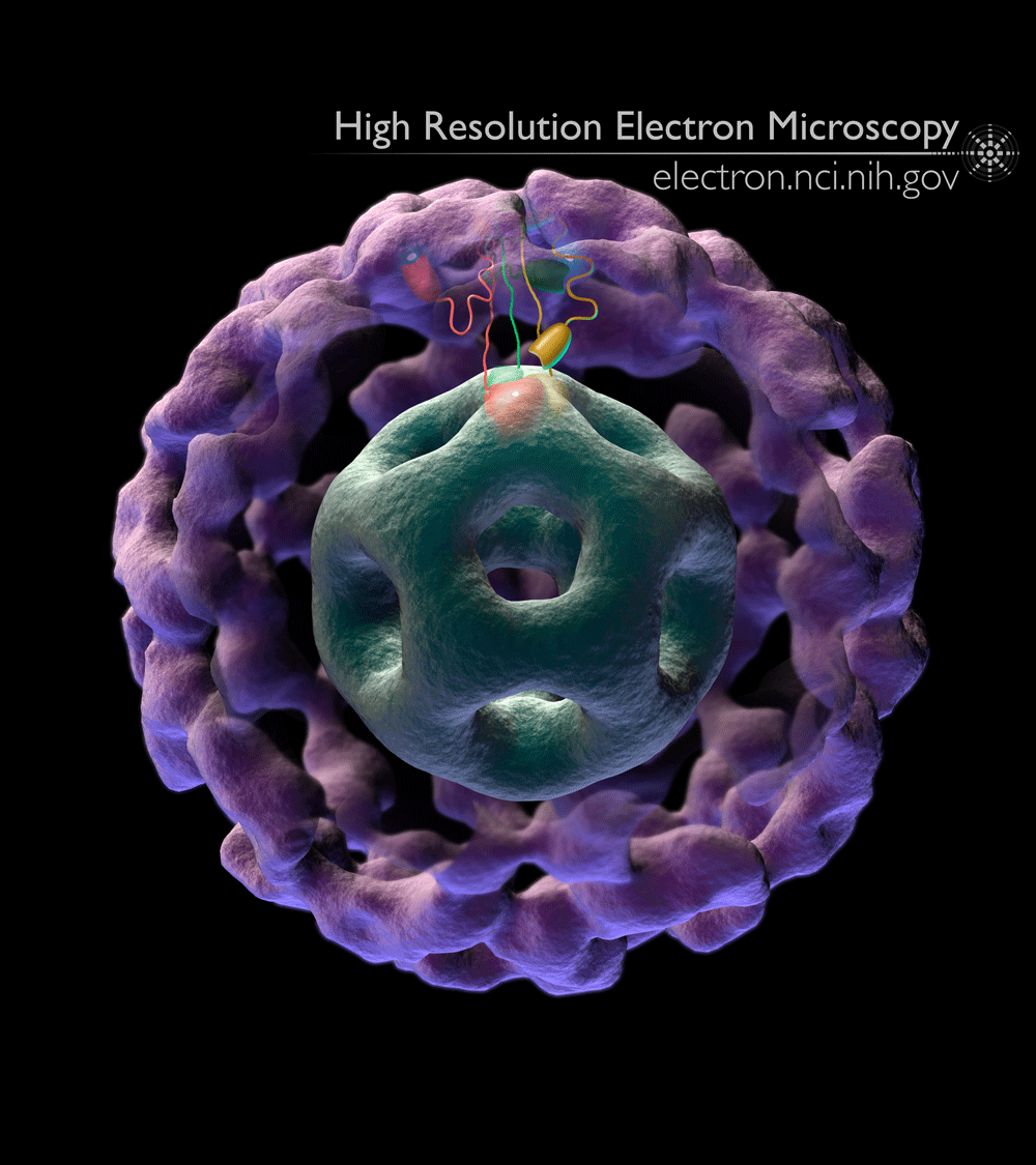
This image shows the inner and outer shells of the large protein machine pyruvate dehydrogenase, which converts pyruvate (derived from sugars like glucose) into acetyl-coA. This is one of many steps necessary for a cell to generate energy, and also provides the building blocks for a variety of molecules that are recycled and reused by the cell.
The image of the pyruvate dehydrogenase enzyme shown here was derived directly from cryo-electron microscopic data. We used a technique called cryo-electron microscopy to image thousands of copies of purified pyruvate dehydrogenase enzyme complexes. In this technique, electrons are shot through a very cold (about – 200C) protein sample towards a detector, which picks up a “shadow” of each protein molecule. Using advanced computer algorithms that can align and analyze thousands of images, these “shadow” snapshots can be used to create a 3D structure of the molecule.
Our video demonstrating pyruvate dehydrogenase at work was featured in the NIH Director’s Blog in 2013.
Our publications on this and related structures of pyruvate dehydrogenase complexes were published in 2002-2008. This structure was originally published in 2002.
Full-size image: Download (0.3MB)
Related Reference: Milne JL, Shi D, et al. Molecular architecture and mechanism of an icosahedral pyruvate dehydrogenase complex: a multifunctional catalytic machine. EMBO J. 2002 Nov 1;21(21):5587-98.